

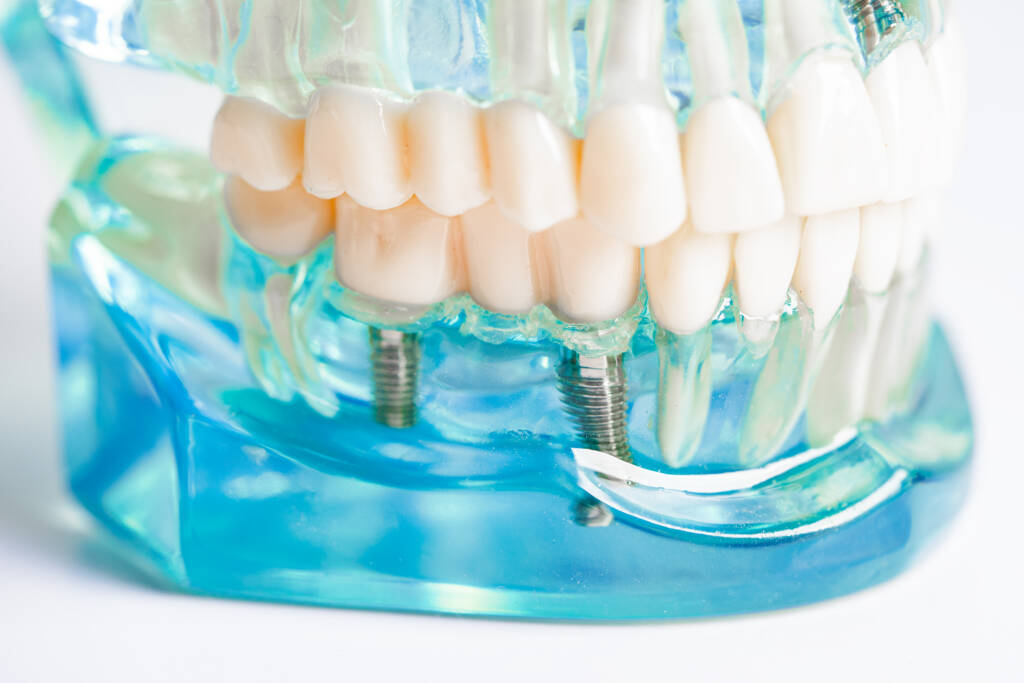
What is Oral Diagnosis?
The word “diagnosis” comes from Greek and means identifying diseases caused by objective and subjective symptoms. Oral diagnosis, on the other hand, is the process of making a diagnosis following the examination of X-rays of the patient’s mouth and a detailed intraoral examination. The primary goal at this stage is to identify the source of the patient’s primary complaint. Furthermore, the patient’s current condition should be considered holistically, identifying not only existing but also potential problem areas that may cause future problems. This allows early diagnosis and preventative and preventive treatments to prevent further problems and make treatment both simpler and more cost-effective.
RADIOLOGY
Panoramic Radiographs
Panoramic radiographs are an extraoral imaging technique that allows for the examination of all existing teeth, impacted teeth, surrounding bone, the entire jawbone, physiological and pathological spaces in the oral shadow, and joints in a single image. It is used particularly in general oral examinations for follow-up purposes, as well as in simple surgical procedures such as extraction of impacted teeth, resection, small cysts, or placement of a small number of implants. Imaging all teeth at once reduces patient radiation exposure compared to serial periapical imaging and allows the physician to make a comprehensive assessment.
Digital Radiography (RVG)
Negative radiology techniques used in the past had several disadvantages, including the time required during the development phase, the chemicals in the development solution, and the environmental impact of lead in the X-ray film. Furthermore, films could easily be damaged with this technique.
This allows X-ray images to be viewed instantly on a computer screen, and the images can be adjusted for color, magnified, reduced, and measured. This technique significantly reduces patient radiation exposure. The ability to store images in the patient’s file or share them online when necessary is a significant advantage.
Computed Tomography (CT)
This three-dimensional tomographic imaging technique is used in cases where traditional intraoral and extraoral imaging techniques are inadequate, such as large cysts, tumors, and cases where multiple implants are planned. Unlike other two-dimensional techniques, this method allows cross-sections to be taken from the oral cavity, allowing the relationship of teeth or pathological lesions to surrounding tissues to be examined in three planes. Horizontal bone thickness, the location of sinus cavities, and the path of blood vessels and nerves can be precisely determined, especially in cases where multiple implants will be placed. Another advantage of this method is that, in combination with rapid prototyping, it allows for the preparation of study models or guide plates for the desired areas prior to surgery. This minimizes the risks associated with surgical procedures.